
Further Your QEEG Knowledge
Advantages of getting your brain mapped
Most people’s problems are beyond their thinking mind and are seen on QEEG brain maps
Discover the power of QEEG Brain Mapping
Determine the root causes of your symptoms
Discover if areas of your brain are working too hard or not enough
QEEG’s help determine if you are taking the right medications
Learn More About QEEG Brain Mapping Solutions
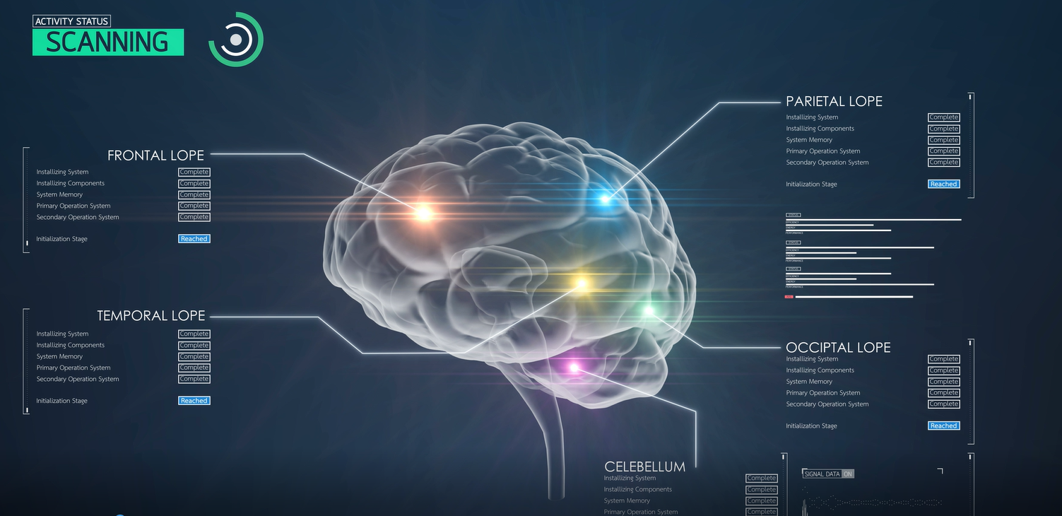
The Electroencephlograph (EEG) is a device that measures electrical-chemical reactions of neurons across the scalp of the cerebral cortex (outer brain). Tiny cup-like sensors capture the signal which is then amplified by the EEG machine. There are 19 sites that are universally accepted, allowing fellow doctors and clinicians to discuss specific regions and ensure everybody is on the same page. The “Q” in QEEG is for the quantitative analysis performed to the raw EEG data collected. Psychologists and Neurotherapists utilize additional software programs to identify areas of the brain that are under or over-activated, and look for correlations of these brain patterns that have been documented in psychological and/or behavioral concerns.
“Many clients are confused by centers that claim to offer ‘QEEG’ Brain Mapping, but in actuality are doing a ‘mini-Q’ Brain Map. Just to be clear, there is a tremendous difference between the two.”
Let us begin by offering a simple explanation of QEEG Brain Mapping. QEEG Brain Mapping, or to be more precise, Quantitative Electroencephalogram, is a procedure that measures and records neural activities of neurons or brain cells in the brain simultaneously from at least 19 locations. It is important that the activities are measured simultaneously, so that we can understand precisely how the brain works as a whole. Studies have continually shown that a minimum of 19 locations is required to accurately map our brain function. More is better but anything less than the desirable 19 locations will not provide sufficient resolution. This is why QEEG Brain Mapping is still considered the “Gold Standard” within the industry.
The Full QEEG (Gold Standard)
19-Channel Brainwave Assessment
The EEG machine records all 19 channels of brainwave activity simultaneously
Full QEEG maps have greater reliability and recorded duration of data
Normative database analysis of all 19 channels
Connectivity of the entire brain analysis demonstrate whole brain connectivity
LORETA inner brain reports are produced
The "Mini-Q"
Brain Map Procedure
The EEG machine only allows limited recording of 2 or 4 channels at a time
Sensors are moved around the head to capture data in 10-12 sites
Normative databases between only 2-4 channels
Connectivity of the entire brain analysis is not possible
LORETA imaging is not possible
What Else Should I Know?

It is important to take note that the mini-Q measures only 2 to 4 sites on the head at a time, while a full QEEG measures at least 19 sites on the head simultaneously. So for a mini-Q to measure 19 sites, it has to be repeated 5 times in sequence if a 4 channel measurement device is used. It would have to be repeated 10 times if a 2 channel measurement device is used. The problem with such sequential measurement is that while you are recording 4 sites, you do not have a clue what is happening at the other 15 or more sites. To say the least, this can give a very misleading picture of what’s going on in the brain.
In a full QEEG, at least 19 sites on the head are measured and recorded simultaneously. This simultaneous measurement is crucial as it allows us to have a good idea on how we use our brain and what goes on when we are engaged on a task. For example: If one location of the brain shows excessive neural activities while on task, we can assess if the other 18 or more sites are affected by this excessive activities. This cannot be done with the mini-Q.
For some reason, the mini-Q measures EEG data only for 1 minute (60 sec). This amount of data is too little to do a rigorous statistical analysis. This is because the recording will usually contain noise, interferences or signals which are not EEG or brain waves, such as eye blinks or movements. These non-EEG signals must be removed before any analysis can be done to assess brain function. Having these contaminants will distort the analysis and lead to a wrong conclusion. So with 60 seconds of raw data, the amount of left data after removal of the contaminants can be insufficient for a good analysis. This can result in arriving at a wrong conclusion.
